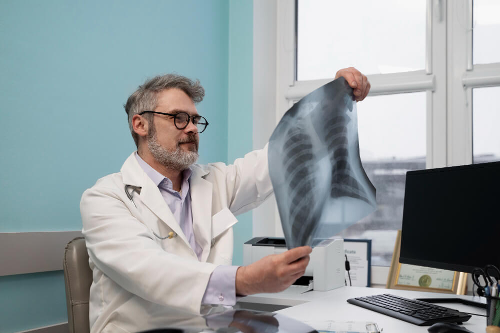
X-Ray Services: A Vital Tool for Fracture Diagnosis
X-rays are a type of electromagnetic radiation that can pass through soft tissues but is absorbed by denser substances like bone. This property makes X-rays an indispensable tool for diagnosing bone fractures and other musculoskeletal injuries.
How X-Ray Technology Works
X-ray machines produce X-rays, which are directed at the body part being examined. The X-rays pass through the body and are captured on a detector. Dense structures, such as bones, absorb more X-rays than soft tissues, resulting in a white image on the film or digital detector.
The Role of X-Rays in Fracture Diagnosis
X-rays are the primary diagnostic tool for detecting bone fractures. They provide clear images of bone structures, allowing healthcare professionals to identify:
- Simple fractures: A clean break in the bone.
- Compound fractures: A fracture where the bone breaks through the skin.
- Stress fractures: Tiny cracks in the bone caused by repetitive stress.
- Growth plate injuries: Fractures that occur in the growth plates of children’s bones.
The X-Ray Procedure
An X-ray procedure is typically quick and painless. Here’s a general overview of the process:
- Preparation: The patient is asked to remove any jewelry or clothing that may interfere with the X-ray image.
- Positioning: The patient is positioned on an X-ray table, ensuring that the affected body part is aligned correctly.
- Image Capture: The X-ray technician takes the image, which may involve multiple exposures from different angles.
- Image Review: The radiologist reviews the images to identify any fractures or other abnormalities.
Interpreting X-Ray Images

Radiologists, highly trained medical professionals, interpret X-ray images to identify fractures and other abnormalities. They look for specific signs of a fracture, such as:
- A break in the bone: A clear line or gap in the bone.
- Displaced bone fragments: Pieces of bone that are out of alignment.
- Swelling or inflammation: Soft tissue swelling around the fracture site.
- Loss of bone density: In cases of osteoporosis or other bone diseases.
Advanced X-Ray Techniques
In addition to traditional X-rays, advanced imaging techniques can provide more detailed information about bone fractures:
- Fluoroscopy: This technique uses real-time X-ray imaging to visualize the movement of bones and joints.
- CT Scan: A CT scan combines multiple X-ray images to create cross-sectional images of the body, providing a more detailed view of bone structures.
- Bone Mineral Density (BMD) Scan: This specialized X-ray measures bone density to assess the risk of osteoporosis.
The Importance of Early Diagnosis and Treatment
Early diagnosis of fractures is crucial for proper treatment and healing. Prompt medical attention can help prevent complications, such as infection or delayed healing. Treatment options for fractures may include:
- Immobilization: Using casts, splints, or braces to immobilize the injured area.
- Surgery: In some cases, surgery may be necessary to repair the fracture, especially for complex fractures or fractures that do not heal properly.
- Physical Therapy: Physical therapy can help restore strength, flexibility, and range of motion.
Conclusion
X-ray technology plays a vital role in the diagnosis and treatment of fractures. By providing clear and detailed images of bone structures, X-rays help healthcare providers make accurate diagnoses and develop effective treatment plans. If you experience any symptoms of a fracture, such as pain, swelling, or difficulty moving, it is important to seek medical attention promptly.
Contact our clinic’s X-ray services for fast and accurate diagnosis of fractures (469) 200-5974 or visit us https://scclittleelm.com/

